Complete Summary and Solutions for Cellular Organelles – NCERT Class XI Biotechnology, Chapter 2 – Structure, Functions, Mechanisms, Exercises
Comprehensive summary and explanation of Chapter 2 'Cellular Organelles' from the NCERT Class XI Biotechnology textbook, covering structure, classification, functions, mechanisms of cellular transport, major organelles, and answers to all textbook questions.
Cellular Organelles: Class 11 NCERT Chapter 2 - Ultimate Study Guide, Notes, Questions, Quiz 2025
Cellular Organelles
Chapter 2: Biotechnology - Ultimate Study Guide | NCERT Class 11 Notes, Questions, Examples & Quiz 2025
Full Chapter Summary & Detailed Notes - Cellular Organelles Class 11 NCERT
Overview & Key Concepts
Chapter Goal: Understand structure and functions of prokaryotic and eukaryotic cells, focusing on organelles. Exam Focus: Fluid mosaic model, transport mechanisms, cell wall differences, endomembrane system. 2025 Updates: Emphasis on dynamic nature of membranes, role in cellular processes. Fun Fact: Cells perform multitasking like digestion and protein synthesis simultaneously. Core Idea: Cells as basic units with specialized organelles. Real-World: Organelle dysfunction links to diseases like mitochondrial disorders. Ties: Builds on cell theory; leads to biomolecules (Ch3). Expanded: All subtopics covered point-wise with diagram descriptions for visual learning.
Wider Scope: From prokaryotic simplicity to eukaryotic complexity; common components like plasma membrane.
Expanded Content: Detailed on all 12 sections (2.1-2.12), including transport, walls, systems, powerhouses, factories, skeletons, motility, control centers.
2.1 Plasma Membrane
Structure & Function: Selective boundary of cytoplasm, guarded by extracellular matrix; maintains cell identity and relations with environment.
Historical Insight: Chemical composition (lipids ~40%, proteins ~52% in RBCs) understood post-electron microscope; Edwin Gorter & F. Grendel (1925) proposed bilayer from RBC lipids covering surface twice.
Fluid Mosaic Model (Singer & Nicolson, 1972): Quasi-fluid lipid bilayer with embedded globular proteins; dynamic, allows lateral diffusion of lipids/proteins.
Components: Phospholipids (hydrophilic heads outward, hydrophobic tails inward); cholesterol for fluidity; peripheral proteins (signaling, superficial); integral proteins (transmembrane channels, most abundant).
Prokaryotic Similarity: Structurally akin to eukaryotic membranes.
Special Structures: Mesosomes (extensions as vesicles/tubules/lamellae) increase surface area in prokaryotes.
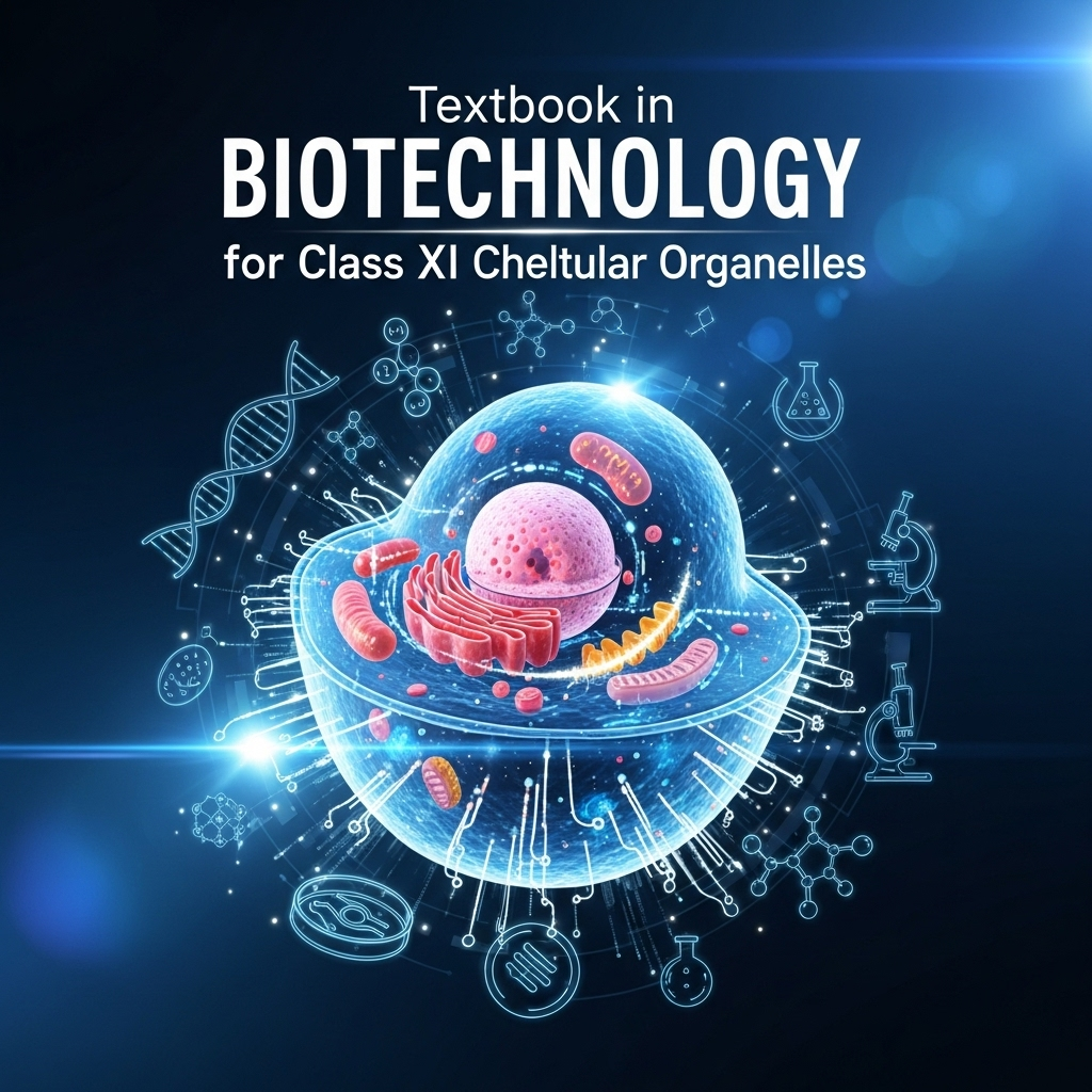
Fig. 2.1: Schematic Diagram of Fluid Mosaic Model (Description)
Bilayer with hydrophilic heads outside/inside, hydrophobic tails in middle; embedded integral/peripheral proteins, cholesterol, glycoproteins/glycolipids on outer surface; shows channel proteins and quasifluid state.
Box 1: Gorter & Grendel Experiment (Description)
RBCs washed/extracted with acetone; lipids formed two-molecule layer covering cell surface; electron micrograph shows 'railroad track' (dense lines for heads, light for tails); proposed bilayer over monolayer.
(a) Cellulose: Linear β-D-glucose chains via β1→4 glycosidic bonds. (b) Chitin: Linear N-acetylglucosamine chains via β1→4 glycosidic bonds.
2.3 Endomembrane System
Overview: Coordinated membrane-bound organelles for protein/lipid synthesis, processing, packaging, transport; includes ER, Golgi, lysosomes, vacuoles.
Tip: Group by prokary/eukary; examples for recall. Depth: Functions link to processes. Errors: Confuse RER/SER. Historical: Gorter 1925 bilayer. Interlinks: Biomolecules Ch3. Advanced: Endosymbiosis. Real-Life: Cilia defects in respiratory diseases. Graphs: Fig 2.1 model. Coherent: Structure → Function → Transport. For easy learning: Flashcard per term.
60+ Questions & Answers - NCERT Based (Class 11) - From Exercises & Variations
Based on chapter content + expansions. Part A: 10 (1 mark short, one line each), Part B: 10 (4 marks medium, five lines each), Part C: 10 (6 marks long, eight lines each). Answers point-wise, step-by-step for marks. Easy learning: Structured, concise.
Part A: 1 Mark Questions (10 Qs - Short from Content)
1. Who proposed the fluid mosaic model of plasma membrane?
1 Mark Answer: Singer and Nicolson in 1972.
2. What is the major lipid in plasma membrane?
1 Mark Answer: Phospholipids.
3. Name the transport requiring ATP against gradient.
1 Mark Answer: Active transport.
4. What is the bacterial cell wall component?
1 Mark Answer: Peptidoglycan.
5. Differentiate Gram-positive and Gram-negative walls.
1 Mark Answer: Gram-positive thick peptidoglycan; Gram-negative thin with outer membrane.
6. What is the plant cell wall polymer?
1 Mark Answer: Cellulose.
7. Name the endomembrane system components.
1 Mark Answer: ER, Golgi, lysosomes, vacuoles.
8. What is the function of mitochondria?
1 Mark Answer: ATP production.
9. What is the 9+2 structure in?
1 Mark Answer: Cilia and flagella.
10. What organizes spindle in animal cells?
1 Mark Answer: Centrioles.
Part B: 4 Marks Questions (10 Qs - Medium, Exactly 5 Lines Each)
1. Describe the fluid mosaic model.
4 Marks Answer:
Proposed by Singer and Nicolson in 1972.
Lipid bilayer with embedded proteins in mosaic pattern.
Quasifluid state allows lateral diffusion.
Phospholipids: Hydrophilic heads out, tails in.
Proteins: Peripheral (signaling), integral (channels).
2. Explain passive and facilitated transport.
4 Marks Answer:
Passive: No energy, along gradient via diffusion/osmosis.
Limited to small uncharged molecules.
Facilitated: Carrier/channel proteins for larger/charged (e.g., glucose).
Aquaporins: Water-specific channels.
Ion channels: Gated in muscle/nerve.
3. Describe Na+-K+ pump mechanism.
4 Marks Answer:
Active transport using ATP.
Pumps 3 Na+ out, 2 K+ in against gradient.
Maintains -60mV potential.
High Na+ outside, high K+ inside.
Essential for nerve/muscle function.
4. Differentiate prokaryotic cell walls.
4 Marks Answer:
Gram-positive: Thick peptidoglycan, teichoic acid, single membrane.
2 attempts on CBSE Class 11 Annual Assessment quizzes
f scored 4/10 on Accountancy (Class 11) Subhash scored 1/10 on Sets and Venn Operations f scored 4/10 on Accountancy (Class 11) Subhash scored 1/10 on Sets and Venn Operations
#1
Accountancy (Class 11) Practice Quiz | CBSE Class 11 Annual Assessment
10 Qs · ~10 min
#2
Sets and Venn Operations Fundamentals — Free CBSE Class 11 Annual Assessment Quiz
10 Qs · ~10 min
#3
Class 11 English — The Tale of Melon City (Practice Quiz)
10 Qs · ~10 min
#4
Class 11 English — Birth (Practice Quiz)
10 Qs · ~10 min
#5
Class 11 English — Mother's Day (Practice Quiz)
10 Qs · ~10 min
#6
Class 11 English — The Address (Practice Quiz)
10 Qs · ~10 min
#7
Class 11 English — The Summer of the Beautiful White Horse (Practice Quiz)
10 Qs · ~10 min
#8
Class 11 English — Father to Son (Practice Quiz)
10 Qs · ~10 min
#9
Class 11 English — Silk Road (Practice Quiz)
10 Qs · ~10 min
#10
Class 11 English — The Adventure (Practice Quiz)
10 Qs · ~10 min
#11
Class 11 English — Childhood (Practice Quiz)
10 Qs · ~10 min
#12
Class 11 English — The Ailing Planet: the Green Movement's Role (Practice Quiz)
10 Qs · ~10 min
#13
Class 11 English — The Voice of the Rain (Practice Quiz)
10 Qs · ~10 min
#14
Class 11 English — The Laburnum Top (Practice Quiz)
10 Qs · ~10 min
#15
Class 11 English — Discovering Tut: the Saga Continues (Practice Quiz)
10 Qs · ~10 min
#16
Class 11 English — We're Not Afraid to Die... if We Can All Be Together (Practice Quiz)
10 Qs · ~10 min
#17
Class 11 English — A Photograph (Practice Quiz)
10 Qs · ~10 min
#18
Class 11 English — The Portrait of a Lady (Practice Quiz)
10 Qs · ~10 min
#19
Class 11 Psychology — Motivation and Emotion (Practice Quiz)
10 Qs · ~10 min
#20
Class 11 Psychology — Thinking (Practice Quiz)
10 Qs · ~10 min
#21
Class 11 Psychology — Human Memory (Practice Quiz)
10 Qs · ~10 min
#22
Class 11 Psychology — Learning (Practice Quiz)
10 Qs · ~10 min
#23
Class 11 Psychology — Sensory, Attentional and Perceptual Processes (Practice Quiz)
10 Qs · ~10 min
#24
Class 11 Psychology — Human Development (Practice Quiz)
10 Qs · ~10 min
#25
Class 11 Psychology — Methods of Enquiry in Psychology (Practice Quiz)
10 Qs · ~10 min
#26
Class 11 Psychology — What is Psychology? (Practice Quiz)
10 Qs · ~10 min
#27
Class 11 Sociology — Indian Sociologists (Practice Quiz)
10 Qs · ~10 min
#28
Class 11 Sociology — Introducing Western Sociologists (Practice Quiz)
10 Qs · ~10 min
#29
Class 11 Sociology — Environment and Society (Practice Quiz)
10 Qs · ~10 min
#30
Class 11 Sociology — Social Change and Social Order in Rural and Urban Society (Practice Quiz)
10 Qs · ~10 min
#31
Class 11 Sociology — Social Structure, Stratification and Social Processes in Society (Practice Quiz)
10 Qs · ~10 min
#32
Class 11 Sociology — Doing Sociology: Research Methods (Practice Quiz)
10 Qs · ~10 min
#33
Class 11 Sociology — Culture and Socialisation (Practice Quiz)
10 Qs · ~10 min
#34
Class 11 Sociology — Understanding Social Institutions (Practice Quiz)
10 Qs · ~10 min
#35
Class 11 Sociology — Terms, Concepts and Their Use in Sociology (Practice Quiz)
10 Qs · ~10 min
#36
Class 11 Sociology — Sociology and Society (Practice Quiz)
10 Qs · ~10 min
#37
Class 11 Political Science — The Philosophy of the Constitution (Practice Quiz)
10 Qs · ~10 min
#38
Class 11 Political Science — Constitution as a Living Document (Practice Quiz)
10 Qs · ~10 min
#39
Class 11 Political Science — Local Governments (Practice Quiz)
10 Qs · ~10 min
#40
Class 11 Political Science — Federalism (Practice Quiz)
10 Qs · ~10 min
#41
Class 11 Political Science — Judiciary (Practice Quiz)
10 Qs · ~10 min
#42
Class 11 Political Science — Legislature (Practice Quiz)
10 Qs · ~10 min
#43
Class 11 Political Science — Executive (Practice Quiz)
10 Qs · ~10 min
#44
Class 11 Political Science — Election and Representation (Practice Quiz)
10 Qs · ~10 min
#45
Class 11 Political Science — Rights in the Indian Constitution (Practice Quiz)
10 Qs · ~10 min
#46
Class 11 Political Science — Constitution: Why and How? (Practice Quiz)
10 Qs · ~10 min
#47
Class 11 Political Science — Secularism (Practice Quiz)
10 Qs · ~10 min
#48
Class 11 Political Science — Nationalism (Practice Quiz)
10 Qs · ~10 min
#49
Class 11 Political Science — Citizenship (Practice Quiz)
10 Qs · ~10 min
#50
Class 11 Political Science — Rights (Practice Quiz)
10 Qs · ~10 min
#51
Class 11 Political Science — Social Justice (Practice Quiz)
10 Qs · ~10 min
#52
Class 11 Political Science — Equality (Practice Quiz)
10 Qs · ~10 min
#53
Class 11 Political Science — Freedom (Practice Quiz)
10 Qs · ~10 min
#54
Class 11 Political Science — Political Theory: An Introduction (Practice Quiz)
10 Qs · ~10 min
#55
Class 11 Geography — Natural Hazards and Disasters (Practice Quiz)
10 Qs · ~10 min
#56
Class 11 Geography — Natural Vegetation (Practice Quiz)
10 Qs · ~10 min
#57
Class 11 Geography — Climate (Practice Quiz)
10 Qs · ~10 min
#58
Class 11 Geography — Drainage System (Practice Quiz)
10 Qs · ~10 min
#59
Class 11 Geography — Structure and Physiography (Practice Quiz)
10 Qs · ~10 min
#60
Class 11 Geography — India – Location (Practice Quiz)
10 Qs · ~10 min
#61
Class 11 Geography — Biodiversity and Conservation (Practice Quiz)
10 Qs · ~10 min
#62
Class 11 Geography — Movements of Ocean Water (Practice Quiz)
10 Qs · ~10 min
#63
Class 11 Geography — Water (Oceans) (Practice Quiz)
10 Qs · ~10 min
#64
Class 11 Geography — World Climate and Climate Change (Practice Quiz)
10 Qs · ~10 min
#65
Class 11 Geography — Water in the Atmosphere (Practice Quiz)
10 Qs · ~10 min
#66
Class 11 Geography — Atmospheric Circulation and Weather Systems (Practice Quiz)
10 Qs · ~10 min
#67
Class 11 Geography — Solar Radiation, Heat Balance and Temperature (Practice Quiz)
10 Qs · ~10 min
#68
Class 11 Geography — Composition and Structure of Atmosphere (Practice Quiz)
10 Qs · ~10 min
#69
Class 11 Geography — Landforms and their Evolution (Practice Quiz)
10 Qs · ~10 min
#70
Class 11 Geography — Geomorphic Processes (Practice Quiz)
10 Qs · ~10 min
#71
Class 11 Geography — Distribution of Oceans and Continents (Practice Quiz)
10 Qs · ~10 min
#72
Class 11 Geography — Interior of the Earth (Practice Quiz)
10 Qs · ~10 min
#73
Class 11 Geography — The Origin and Evolution of the Earth (Practice Quiz)
10 Qs · ~10 min
#74
Class 11 Geography — Geography as a Discipline (Practice Quiz)
10 Qs · ~10 min
#75
Class 11 History — Paths to Modernisation (Practice Quiz)
10 Qs · ~10 min
#76
Class 11 History — Displacing Indigenous Peoples (Practice Quiz)
10 Qs · ~10 min
#77
Class 11 History — Changing Cultural Traditions (Practice Quiz)
10 Qs · ~10 min
#78
Class 11 History — The Three Orders (Practice Quiz)
10 Qs · ~10 min
#79
Class 11 History — Nomadic Empires (Practice Quiz)
10 Qs · ~10 min
#80
Class 11 History — An Empire Across Three Continents (Practice Quiz)
10 Qs · ~10 min
#81
Class 11 History — Writing and City Life (Practice Quiz)
10 Qs · ~10 min
#82
Class 11 Economics — Comparative Development Experiences of India and its Neighbours (Practice Quiz)
10 Qs · ~10 min
#83
Class 11 Economics — Environment and Sustainable Development (Practice Quiz)
10 Qs · ~10 min
#84
Class 11 Economics — Employment: Growth, Informalisation and Other Issues (Practice Quiz)
10 Qs · ~10 min
#85
Class 11 Economics — Rural Development (Practice Quiz)
10 Qs · ~10 min
#86
Class 11 Economics — Human Capital Formation in India (Practice Quiz)
10 Qs · ~10 min
#87
Class 11 Economics — Liberalisation, Privatisation and Globalisation: An Appraisal (Practice Quiz)
10 Qs · ~10 min
#88
Class 11 Economics — Indian Economy 1950-1990 (Practice Quiz)
10 Qs · ~10 min
#89
Class 11 Economics — Indian Economy on the Eve of Independence (Practice Quiz)
10 Qs · ~10 min
#90
Class 11 Economics — Use of Statistical Tools (Practice Quiz)
10 Qs · ~10 min
#91
Class 11 Economics — Index Numbers (Practice Quiz)
10 Qs · ~10 min
#92
Class 11 Economics — Correlation (Practice Quiz)
10 Qs · ~10 min
#93
Class 11 Economics — Measures of Central Tendency (Practice Quiz)
10 Qs · ~10 min
#94
Class 11 Economics — Presentation of Data (Practice Quiz)
10 Qs · ~10 min
#95
Class 11 Economics — Organisation of Data (Practice Quiz)
10 Qs · ~10 min
#96
Class 11 Economics — Collection of Data (Practice Quiz)
10 Qs · ~10 min
#97
Class 11 Economics — Introduction (Practice Quiz)
10 Qs · ~10 min
#98
Class 11 Business Studies — International Business (Practice Quiz)
10 Qs · ~10 min
#99
Class 11 Business Studies — Internal Trade (Practice Quiz)
10 Qs · ~10 min
#100
Class 11 Business Studies — MSME and Business Entrepreneurship (Practice Quiz)
10 Qs · ~10 min
#101
Class 11 Business Studies — Sources of Business Finance (Practice Quiz)
10 Qs · ~10 min
#102
Class 11 Business Studies — Formation of a Company (Practice Quiz)
10 Qs · ~10 min
#103
Class 11 Business Studies — Social Responsibilities of Business and Business Ethics (Practice Quiz)
10 Qs · ~10 min
#104
Class 11 Business Studies — Emerging Modes of Business (Practice Quiz)
10 Qs · ~10 min
#105
Class 11 Business Studies — Business Services (Practice Quiz)
10 Qs · ~10 min
#106
Class 11 Business Studies — Private, Public and Global Enterprises (Practice Quiz)
10 Qs · ~10 min
#107
Class 11 Business Studies — Forms of Business Organisation (Practice Quiz)
10 Qs · ~10 min
#108
Class 11 Business Studies — Business, Trade and Commerce (Practice Quiz)
10 Qs · ~10 min
#109
Class 11 Accountancy — Financial Statements - II (Practice Quiz)
10 Qs · ~10 min
#110
Class 11 Accountancy — Financial Statements - I (Practice Quiz)
10 Qs · ~10 min
#111
Class 11 Accountancy — Depreciation, Provisions and Reserves (Practice Quiz)
10 Qs · ~10 min
#112
Class 11 Accountancy — Trial Balance and Rectification of Errors (Practice Quiz)
10 Qs · ~10 min
#113
Class 11 Accountancy — Bank Reconciliation Statement (Practice Quiz)
10 Qs · ~10 min
#114
Class 11 Accountancy — Recording of Transactions - II (Practice Quiz)
10 Qs · ~10 min
#115
Class 11 Accountancy — Recording of Transactions - I (Practice Quiz)
10 Qs · ~10 min
#116
Class 11 Accountancy — Theory Base of Accounting (Practice Quiz)
10 Qs · ~10 min
#117
Class 11 Accountancy — Introduction to Accounting (Practice Quiz)
10 Qs · ~10 min
#118
Class 11 Maths — Probability (Practice Quiz)
10 Qs · ~10 min
#119
Class 11 Maths — Statistics (Practice Quiz)
10 Qs · ~10 min
#120
Class 11 Maths — Limits and Derivatives (Practice Quiz)
10 Qs · ~10 min
#121
Class 11 Maths — Introduction to Three Dimensional Geometry (Practice Quiz)
10 Qs · ~10 min
#122
Class 11 Maths — Conic Sections (Practice Quiz)
10 Qs · ~10 min
#123
Class 11 Maths — Straight Lines (Practice Quiz)
10 Qs · ~10 min
#124
Class 11 Maths — Sequences and Series (Practice Quiz)
10 Qs · ~10 min
#125
Class 11 Maths — Binomial Theorem (Practice Quiz)
10 Qs · ~10 min
#126
Class 11 Maths — Permutations and Combinations (Practice Quiz)
10 Qs · ~10 min
#127
Class 11 Maths — Linear Inequalities (Practice Quiz)
10 Qs · ~10 min
#128
Class 11 Maths — Complex Numbers and Quadratic Equations (Practice Quiz)
10 Qs · ~10 min
#129
Class 11 Maths — Relations and Functions (Practice Quiz)
10 Qs · ~10 min
#130
Class 11 Maths — Sets (Practice Quiz)
10 Qs · ~10 min
#131
Class 11 Biology — Chemical Coordination and Integration (Practice Quiz)
10 Qs · ~10 min
#132
Class 11 Biology — Neural Control and Coordination (Practice Quiz)
10 Qs · ~10 min
#133
Class 11 Biology — Locomotion and Movement (Practice Quiz)
10 Qs · ~10 min
#134
Class 11 Biology — Excretory Products and their Elimination (Practice Quiz)
10 Qs · ~10 min
#135
Class 11 Biology — Body Fluids and Circulation (Practice Quiz)
10 Qs · ~10 min
#136
Class 11 Biology — Breathing and Exchange of Gases (Practice Quiz)
10 Qs · ~10 min
#137
Class 11 Biology — Plant Growth and Development (Practice Quiz)
10 Qs · ~10 min
#138
Class 11 Biology — Respiration in Plants (Practice Quiz)
10 Qs · ~10 min
#139
Class 11 Biology — Photosynthesis in Higher Plants (Practice Quiz)
10 Qs · ~10 min
#140
Class 11 Biology — Cell Cycle and Cell Division (Practice Quiz)
10 Qs · ~10 min
#141
Class 11 Biology — Biomolecules (Practice Quiz)
10 Qs · ~10 min
#142
Class 11 Biology — Cell: The Unit of Life (Practice Quiz)
10 Qs · ~10 min
#143
Class 11 Biology — Structural Organisation in Animals (Practice Quiz)
10 Qs · ~10 min
#144
Class 11 Biology — Anatomy of Flowering Plants (Practice Quiz)
10 Qs · ~10 min
#145
Class 11 Biology — Morphology of Flowering Plants (Practice Quiz)
10 Qs · ~10 min
#146
Class 11 Biology — Animal Kingdom (Practice Quiz)
10 Qs · ~10 min
#147
Class 11 Biology — Plant Kingdom (Practice Quiz)
10 Qs · ~10 min
#148
Class 11 Biology — Biological Classification (Practice Quiz)
10 Qs · ~10 min
#149
Class 11 Biology — The Living World (Practice Quiz)
10 Qs · ~10 min
#150
Class 11 Chemistry — Hydrocarbons (Practice Quiz)
10 Qs · ~10 min
#151
Class 11 Chemistry — Organic Chemistry – Some Basic Principles and Techniques (Practice Quiz)
10 Qs · ~10 min
#152
Class 11 Chemistry — Redox Reactions (Practice Quiz)
10 Qs · ~10 min
#153
Class 11 Chemistry — Equilibrium (Practice Quiz)
10 Qs · ~10 min
#154
Class 11 Chemistry — Chemical Bonding and Molecular Structure (Practice Quiz)
10 Qs · ~10 min
#155
Class 11 Chemistry — Classification of Elements and Periodicity in Properties (Practice Quiz)
10 Qs · ~10 min
#156
Class 11 Chemistry — Some Basic Concepts of Chemistry (Practice Quiz)
10 Qs · ~10 min
#157
Class 11 Physics — Waves (Practice Quiz)
10 Qs · ~10 min
#158
Class 11 Physics — Oscillations (Practice Quiz)
10 Qs · ~10 min
#159
Class 11 Physics — Kinetic Theory (Practice Quiz)
10 Qs · ~10 min
#160
Class 11 Chemistry — Thermodynamics (Practice Quiz)
10 Qs · ~10 min
#161
Class 11 Physics — Thermal Properties of Matter (Practice Quiz)
10 Qs · ~10 min
#162
Class 11 Physics — Mechanical Properties of Fluids (Practice Quiz)
10 Qs · ~10 min
#163
Class 11 Physics — Mechanical Properties of Solids (Practice Quiz)
10 Qs · ~10 min
#164
Class 11 Physics — Gravitation (Practice Quiz)
10 Qs · ~10 min
#165
Class 11 Physics — Systems of Particles and Rotational Motion (Practice Quiz)
10 Qs · ~10 min
#166
Class 11 Physics — Work, Energy and Power (Practice Quiz)
10 Qs · ~10 min
#167
Class 11 Physics — Motion in a Plane (Practice Quiz)
10 Qs · ~10 min
#168
Class 11 Physics — Motion in a Straight Line (Practice Quiz)
10 Qs · ~10 min
#169
Class 11 Physics — Units and Measurement (Practice Quiz)
10 Qs · ~10 min
#170
Class 11 Maths — Trigonometric Functions (Practice Quiz)
10 Qs · ~10 min
#171
Class 11 Chemistry — Structure of Atom (Practice Quiz)
10 Qs · ~10 min
#172
Class 11 Physics — Laws of Motion (Practice Quiz)
10 Qs · ~10 min
#173
Business Studies (Class 11) Practice Quiz | CBSE Class 11 Annual Assessment
10 Qs · ~10 min
#174
Economics (Class 11) Practice Quiz | CBSE Class 11 Annual Assessment
10 Qs · ~10 min
#175
Humanities Subjects Practice Quiz | CBSE Class 11 Annual Assessment
10 Qs · ~10 min
#176
Motion in a Straight Line Practice Quiz | CBSE Class 11 Annual Assessment
10 Qs · ~10 min
#177
Thermodynamic Processes and Laws Advanced Challenge | CBSE Class 11 Annual Assessment